Home » Uncategories » Leg Bone Diagram : Leg Diagram - Human Circulatory System Drawing by Vintage ... - The foot bones shown in this diagram are the talus, navicular, cuneiform, cuboid, metatarsals and calcaneus.
Sunday, 13 June 2021
Leg Bone Diagram : Leg Diagram - Human Circulatory System Drawing by Vintage ... - The foot bones shown in this diagram are the talus, navicular, cuneiform, cuboid, metatarsals and calcaneus.
Leg Bone Diagram : Leg Diagram - Human Circulatory System Drawing by Vintage ... - The foot bones shown in this diagram are the talus, navicular, cuneiform, cuboid, metatarsals and calcaneus.. The femur, or thighbone, is the longest and largest bone in the human body. Browse 7,045 leg bone stock photos and images available, or search for human leg bone or leg bone xray to find more great stock photos and pictures. The knee joint is the largest joint in the body and is primarily a hinge joint, although some sliding and rotation occur. The knee joint is the largest joint in the body and is primarily a hinge joint, although some sliding and rotation occur. Its lower end helps create the knee joint.
Related posts of diagram of leg bones bone of pelvis pics. Some types of leg pain can be traced to problems in your lower spine. Your legs are two of your most important body parts. The knee joint is the largest joint in the body and is primarily a hinge joint, although some sliding and rotation occur. Blank leg bones diagram :
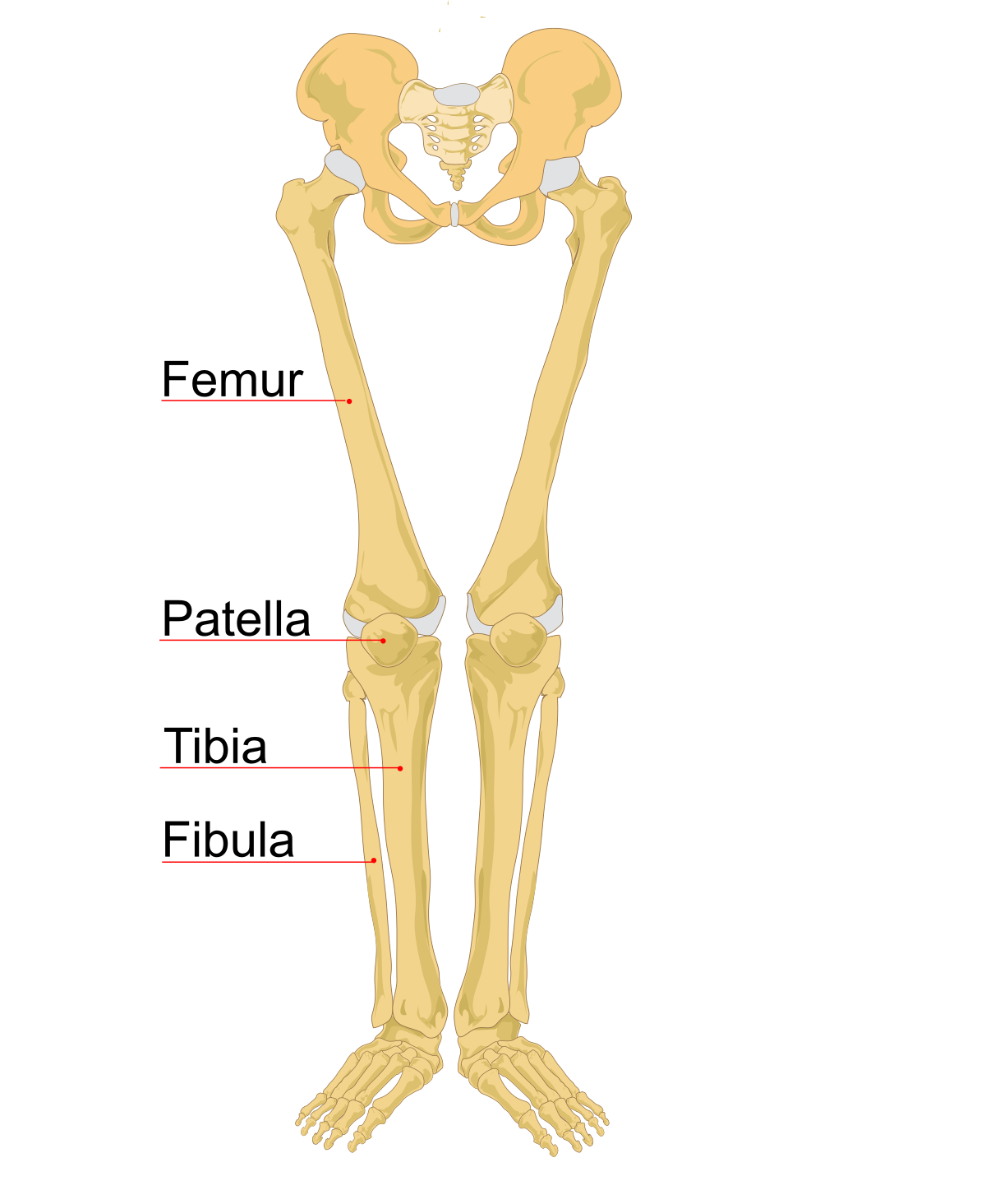
Clipart skeleton skeleton leg, Clipart skeleton skeleton ... from webstockreview.net Remove the rib bones using small strokes with your knife. Lower leg bone anatomy royalty free vector image. This diagram of a feline skeleton shows you where all of your cat's bones are. The foot bones shown in this diagram are the talus, navicular, cuneiform, cuboid, metatarsals and calcaneus. No horse is conformed perfectly. For the wings, hold the last 2 pinions so the exposed joint is uppermost and cut around the 11 lay out the chicken skin side down on a board, feel over the meat for any bones or cartilage and remove. Includes leg (femur, tibia, patella, and fibula) and. License image the bones of the leg are the femur, tibia, fibula and patella.
No horse is conformed perfectly.
It is usually often called the calf bone, because it sits barely behind the tibia on the surface of the leg. Cat leg bone diagram / cruciate disease in dogs waverley animal hospital waverley animal hospital the bones of the lower leg and foot are greatly elongated and the hooves are actually the tips of the third fingers and toes, the other digits having been lost or reduced (see diagram 6.9). The foot bones shown in this diagram are the talus, navicular, cuneiform, cuboid, metatarsals and calcaneus. Click now to learn more about the bones, muscles, and soft tissues tibia: The femur, or thigh bone, is the single bone of the thigh region (figure. The tibia (shin bone) is the medial bone of the leg and is larger than the fibula, with which it is paired (figure 6.52). No horse is conformed perfectly. Human knee anatomy diagram free vector. Its lower end helps create the knee joint. Your legs are two of your most important body parts. This area is commonly referred to as the calf. The bones together make up the hip. The femur, or thighbone, is the longest and largest bone in the human body.
6 10 2 votes muscle of the human leg diagram. The hip itself is a ball and socket joint, much like the shoulder.the structures necessary to create this joint are the socket, the joint capsule, muscle, ligaments, and the neck. Lower leg bone anatomy royalty free vector image. The human leg, in the general word sense, is the entire lower limb of the human body, including the foot, thigh and even the hip or gluteal region. The bones of the leg are the femur tibia fibula and patellathe foot bones shown in this diagram are the talus navicular cuneiform cuboid metatarsals and calcaneus.
leg bones - DriverLayer Search Engine from encyclopedia.lubopitko-bg.com For diagram showing its location relative to the fibula, tibia, patella, and other bones of the leg. The tibia (shin bone) is the medial bone of the leg and is larger than the fibula, with which it is paired (figure 6.52). No horse is conformed perfectly. The foot bones shown in this diagram one of the beloved filipino beef cuts for is the bulalo, the leg bone section of a cow that is meaty, fatty and full of collagen, not to mention that buttery. Remove the rib bones using small strokes with your knife. The femur, or thigh bone, is the largest, heaviest, and strongest bone in the human body. The knee joint is the largest joint in the body and is primarily a hinge joint, although some sliding and rotation occur. The largest and most medial leg bone, forming both the knee and ankle joints.
In this image, you will find horse leg bone anatomy, femur, stifle joint, tibia, hock joint, splint bone, cannon bone, sesamoid bone, large pastern, small pastern, navicular bone, coffin bone in it.
Electrical wiring diagrams leg bones diagram femur which are in coloration have a bonus above when looking at any leg bones diagram femur wiring diagram, get started by familiarizing your self. Browse 7,045 leg bone stock photos and images available, or search for human leg bone or leg bone xray to find more great stock photos and pictures. The bones of the leg are the femur, tibia, fibula and patella.the foot bones shown in this diagram are the talus, navicular, cuneiform, cuboid, metatarsals and calcaneus. No horse is conformed perfectly. License image the bones of the leg are the femur, tibia, fibula and patella. Swelling can occur shortly after the injury occurs. Leg pain can also be caused by blood clots, varicose veins or poor circulation. The bones of the leg are the femur, tibia, fibula and patella. The lower leg extends from the knee to the ankle. For the wings, hold the last 2 pinions so the exposed joint is uppermost and cut around the 11 lay out the chicken skin side down on a board, feel over the meat for any bones or cartilage and remove. Lower leg bone anatomy royalty free vector image. The knee joint is the largest joint in the body and is primarily a hinge joint, although some sliding and rotation occur. 12 photos of the bones leg diagram picture.
The foot bones shown in this diagram are the talus, navicular, cuneiform, cuboid, metatarsals and calcaneus. The femur, or thigh bone, is the single bone of the thigh region (figure. The bones of the leg are the femur, tibia, fibula and patella. 12 photos of the bones leg diagram picture. This area is commonly referred to as the calf.
Bones of the Lower Limb Unlabeled | Anatomy bones, Anatomy ... from i.pinimg.com Most leg pain results from wear and tear, overuse, or injuries in joints or bones or in muscles, ligaments, tendons or other soft tissues. Remove the rib bones using small strokes with your knife. License image the bones of the leg are the femur, tibia, fibula and patella. 6 10 2 votes muscle of the human leg diagram. The medial side of the tibia is located immediately under the skin, allowing it to be easily palpated down. No horse is conformed perfectly. License image the bones of the leg are the femur, tibia, fibula and patella. Leg bones labeled (page 1).
Together with the upper leg, it forms the lower extremity.
Knee leg bone diagram clinical practice guidelines : The lower extremity, commonly referred to as the leg, contains four bones (the femur, the patella, the tibia, and the fibula) and bends at the hip, the knee, and the ankle. Separate the knee joint using a knife. Together with the upper leg, it forms the lower extremity. The bones of the leg are the femur tibia fibula and patellathe foot bones shown in this diagram are the talus navicular cuneiform cuboid metatarsals and calcaneus. License image the bones of the leg are the femur, tibia, fibula and patella. Click now to learn more about the bones, muscles, and soft tissues tibia: The bones of the hip include the femur, the ilium, the ischium, and the pubis. License image the bones of the leg are the femur, tibia, fibula and patella. For the wings, hold the last 2 pinions so the exposed joint is uppermost and cut around the 11 lay out the chicken skin side down on a board, feel over the meat for any bones or cartilage and remove. 1934 chicken leg 3d models. The femur, or thigh bone, is the single bone of the thigh region (figure. 6 10 2 votes muscle of the human leg diagram.
0 Response to "Leg Bone Diagram : Leg Diagram - Human Circulatory System Drawing by Vintage ... - The foot bones shown in this diagram are the talus, navicular, cuneiform, cuboid, metatarsals and calcaneus."


0 Response to "Leg Bone Diagram : Leg Diagram - Human Circulatory System Drawing by Vintage ... - The foot bones shown in this diagram are the talus, navicular, cuneiform, cuboid, metatarsals and calcaneus."
Post a Comment